

Ankle ligament injuries are among the most common musculoskeletal injuries in Australia. They happen in everyday moments: a foot rolling on an uneven kerb during a morning run, a misstep off a gutter, or a landing gone wrong during a weekend basketball game. Most people recognise the immediate discomfort and assume it will resolve on its own with time and rest. Often, that assumption costs them more time in recovery than they bargained for.
Not all ankle sprains are the same, and distinguishing between them from the outset matters. The way an ankle ligament injury is graded at the start of management directly shapes the rehabilitation approach and, ultimately, the outcome. This guide focuses on Grade I ankle ligament tears - the least severe category in clinical grading, but one of the most frequently underestimated.
A Grade I injury still benefits from formal confirmation by a qualified ankle injury specialist. AVA Orthopaedics provides specialist online orthopaedic consultations via secure video for patients anywhere in Australia, including same-day and next-day appointments. No travel required.
The Anatomy of the Ankle Ligament Complex
The ankle joint is formed by the articulation of the tibia, fibula and talus, stabilised by a network of ligamentous structures that provide both mechanical support and proprioceptive feedback. Understanding which part of this complex is injured is central to accurate grading and appropriate management.
The lateral ligament complex is the most commonly injured group, comprising three distinct ligaments. The anterior talofibular ligament (ATFL) is the primary restraint against forward displacement of the talus and inward rotation of the foot, and it is the most frequently torn structure in ankle inversion injuries. The calcaneofibular ligament (CFL) resists inversion stress across both the ankle and the subtalar joint and is often involved alongside the ATFL in higher-grade injuries. The posterior talofibular ligament (PTFL) is the strongest of the three and is rarely injured in isolation.
The medial deltoid ligament complex provides resistance to eversion stress and is less frequently injured. The syndesmotic ligament complex binds the distal tibiofibular joint and is involved in what is known as a high ankle sprain - a mechanically distinct injury with different management requirements. Knowing which structures are affected is fundamental to choosing the right treatment path.
Understanding the Grading System: Grade I, II and III
Ankle ligament injuries are classified on a three-tier scale based on the degree of structural disruption, the presence of joint laxity and the functional impact on the patient.
Grade I: Microscopic Ligament Fibre Disruption
A Grade I injury involves microscopic tearing of individual collagen fibres within the ligament substance, with the overall ligament architecture remaining intact. There is minimal localised swelling and bruising, typically confined to the area directly overlying the injured ligament. No reportable instability from the patient is present. The patient can generally bear weight with mild discomfort, and the joint feels stable during normal walking. Pain is present but manageable.
Grade II: Partial Ligament Tear
A Grade II injury involves a partial macroscopic tear, meaning a significant proportion of ligament fibres have been disrupted at a visible structural level. Moderate swelling, bruising and tenderness are present. Weight-bearing is possible but painful, and functional capacity is reduced.
Grade III: Complete Ligament Rupture
A Grade III injury represents complete disruption of the ligament, with loss of structural continuity. Significant swelling, bruising and tenderness are present. Many patients are unable to bear weight in the acute phase, and functional instability is significant. This category requires formal specialist assessment and MRI review to guide the surgical versus conservative management decision.
Accurate grading by an ankle injury specialist is essential regardless of perceived symptom severity. Grade I injuries can mask concurrent higher-grade tears, osteochondral lesions of the talar dome or avulsion fractures - all of which have direct implications for management if missed early.
What Is an Ankle Ligament Tear Grade I? A Closer Look
A Grade I ankle ligament tear involves stress on the ligament beyond its elastic limit at the collagen fibril level, without producing visible structural failure. Because the gross architecture of the ligament remains intact, the joint is stable and clinical stress testing findings are within normal limits. This is precisely why Grade I injuries are frequently dismissed as minor and left to self-manage.
That dismissal carries real clinical risk as concurrent injuries or differential diagnoses - an osteochondral lesion of the talus, a peroneal tendon injury or a fifth metatarsal avulsion fracture - may go undetected. Without structured rehabilitation, the proprioceptive deficits that accompany even a microscopic ligament injury persist, significantly increasing the likelihood of re-injury and eventual progression to chronic ankle instability. Athletes who return to sport without formal clearance from an ankle injury specialist are at elevated risk of sustaining a higher-grade tear at the same ankle.
Causes and Mechanisms of a Grade I Ankle Ligament Tear
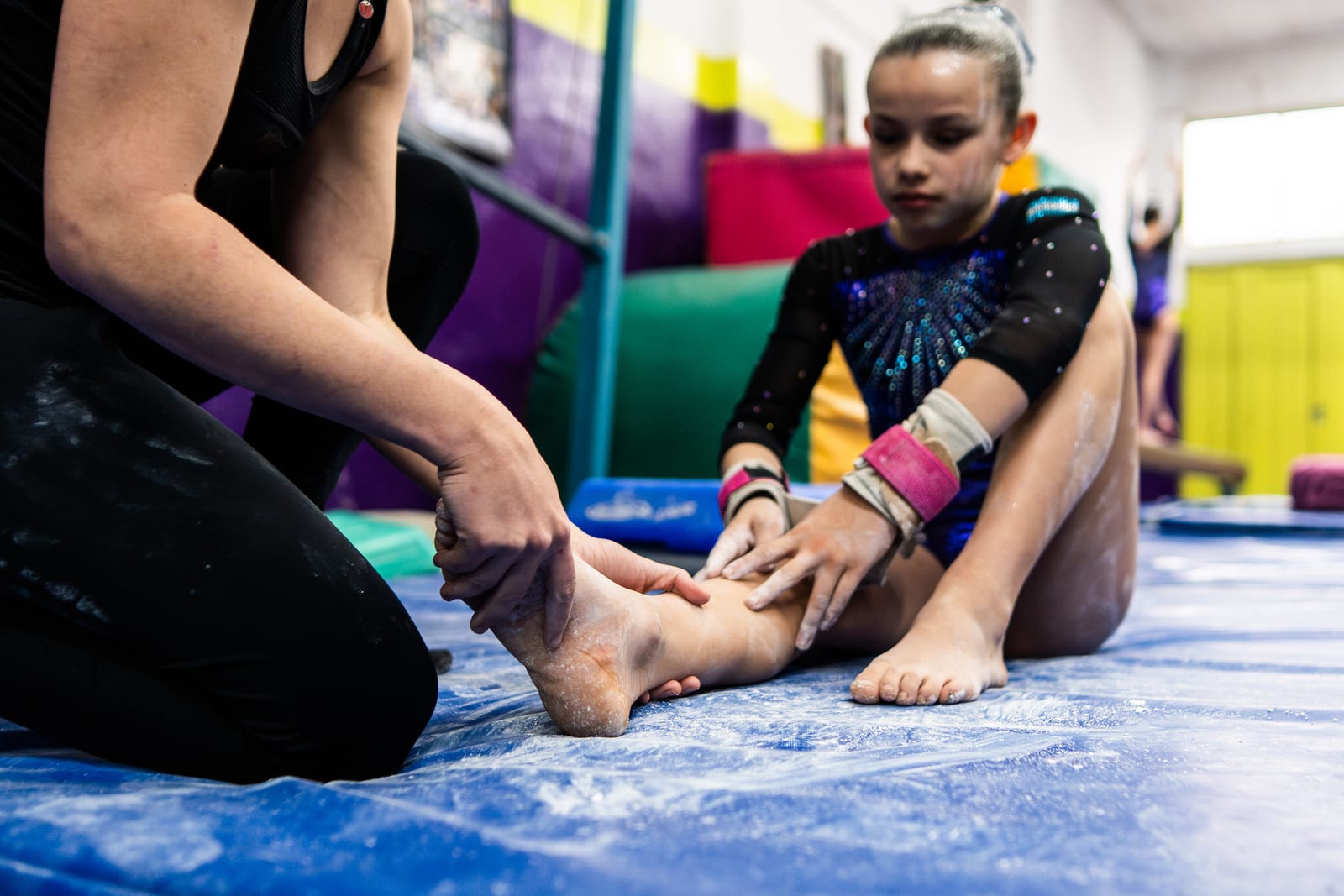
The most common mechanism is inversion combined with plantarflexion - the foot rolling inward and downward under load, placing tensile stress on the lateral ligament complex. Familiar scenarios include landing from a jump, running on uneven terrain, stepping off a kerb awkwardly, or pivoting during team sport.
Several factors increase vulnerability to injury. A prior ankle sprain with incomplete rehabilitation is among the most significant, as residual proprioceptive deficit and mechanical weakness create a lower threshold for re-injury. Reduced ankle dorsiflexion range of motion, peroneal muscle weakness or delayed neuromuscular activation, generalised joint hypermobility, worn or inappropriate footwear and physical fatigue during the latter stages of training or competition all contribute.
A high ankle sprain - involving the syndesmotic ligament complex rather than the lateral ligaments - is produced by a different mechanism: dorsiflexion combined with external rotation of the foot. Distinguishing these two injury patterns is clinically important, as high ankle sprain treatment differs substantially from lateral ligament management and typically involves a longer protected recovery period.
Symptoms of a Grade I Ankle Ligament Tear
A Grade I presentation typically includes localised pain over the anterior talofibular ligament felt immediately at the time of injury, mild swelling developing over the first several hours confined to the lateral ankle, and minimal or no bruising. Tenderness is present on direct assessment of the ATFL and the anterolateral gutter. The patient can bear weight, though discomfort increases on uneven surfaces or during directional changes. There is no subjective sense of instability or giving way.
Mild symptoms should not be equated with a minor injury requiring no further assessment. Consulting an ankle injury specialist can assist you in confirming the injury type, exclude associated pathology and start rehabilitation on the right footing.
How Is a Grade I Ankle Ligament Tear Diagnosed?
Diagnosis begins with a thorough clinical history covering the mechanism of injury, symptom onset, previous ankle injuries and the patient's functional demands. The specialist then reviews any available imaging and can guide the patient through a structured history-based and imaging-supported assessment via secure video consultation.
For imaging, a weight-bearing X-ray guided by the Ottawa Ankle Rules is used to exclude fractures when bony tenderness is present. MRI is not routinely required for Grade I injuries but is indicated when diagnostic uncertainty exists or when symptoms do not resolve as expected.
AVA Orthopaedics specialist consultations allow an orthopaedic specialist to review imaging reports, interpret clinical history and devise a personalised management plan for your ankle injury.
Grade I Ankle Ligament Tear Treatment: A Structured Recovery Plan
Even though a Grade I injury is the least severe category, structured management principles still apply. Outcomes are consistently better with specialist-directed rehabilitation than with unassisted self-management. Physiotherapy greatly assists with rehabilitation guidance.
Acute Phase Management: Days 1 to 5
The PRICE protocol forms the foundation of early management: protection, relative rest, ice application, compression bandaging and elevation. HARM factors should be avoided in the first 72 hours: heat, alcohol, running and massage all increase swelling and impair early tissue recovery. Appropriate analgesia, including paracetamol and anti-inflammatory medications as directed, supports comfort. Early weight-bearing as tolerated is recommended; complete rest is not advised and delays recovery. Taping or bracing provides proprioceptive feedback and confidence during early mobilisation.
Rehabilitation Phase: Days 5 to 21
Rehabilitation focuses on restoring range of motion through ankle alphabet exercises and towel stretches, progressive peroneal and calf strengthening to restore dynamic stabiliser function, and proprioceptive retraining through single-leg balance progressions on stable and then unstable surfaces. Gait retraining normalises walking pattern and eliminates compensatory movement strategies that can place adjacent structures under unnecessary load.
Return to Sport Phase: Weeks 3 to 6
Return to sport should be criteria-based rather than time-based. Sport-specific agility and cutting drills are introduced progressively, with functional performance benchmarks, including single-leg hop testing, figure-of-eight running, and reactive agility tasks, used to confirm readiness. Formal clearance from an orthopaedic specialist or ankle injury doctor before returning to full competition is important. Ongoing proprioceptive training should continue as long-term injury prevention after return to activity.
High Ankle Sprain Treatment: Why It Differs From a Lateral Ligament Injury
The syndesmotic ligament complex binds the distal tibiofibular joint and is a separate structure from the lateral ankle ligaments. A high ankle sprain occurs when external rotation of the foot on a fixed tibia stresses this complex: a mechanism commonly seen in contact sports such as AFL, rugby and soccer.
The clinical presentation of a high ankle sprain differs from a lateral ligament injury in meaningful ways. Pain is felt proximal to the ankle joint rather than over the outer ankle; the squeeze test is positive, and pain is reproduced with external rotation stress. Recovery is typically prolonged compared with lateral ligament injuries. High ankle sprain treatment involves more conservative load management, a longer protected weight-bearing period, and, in cases of complete syndesmotic disruption, surgical stabilisation.
A high ankle sprain misclassified as a routine lateral sprain and undertreated is one of the most common causes of prolonged ankle pain and delayed return to sport. Confirming the diagnosis with an ankle injury specialist at AVA Orthopaedics removes that risk.
Why Online Orthopaedic Specialist Consultation Works for Ankle Ligament Injuries
A common assumption is that an ankle injury must be physically examined in a clinic to receive meaningful specialist input. For ankle ligament injuries, that assumption is not well-founded. The diagnosis is primarily history-based and imaging-supported, and both are well suited to a structured online consultation.
Injury grading is confirmed through a detailed history of the mechanism, the patient's symptom profile, and a review of any available imaging. A structured activity modification plan, bracing recommendation and return-to-sport pathway can be clearly communicated and explained via secure video. Follow-up milestone reviews are easily conducted online, reducing the burden of repeated travel to a clinic - particularly for patients in regional, rural and remote areas of Australia.
AVA Orthopaedics provides specialist orthopaedic consultations via secure video, accessible to anyone in Australia. Same-day and next-day appointments are frequently available, ensuring patients receive timely specialist input at the most critical early stage of recovery.
Book an Online Assessment for Specialist Advice, Treatment Direction and Clarity

Even a mild ankle injury raises legitimate clinical questions. How bad is my ankle sprain? Are there any associated injuries? When is it safe to return to sport? A single consultation with an orthopaedic specialist at AVA Orthopaedics answers those questions with clinical precision, providing a confirmed diagnosis, a structured recovery plan and a clear return-to-sport or daily activities direction.
Before your consultation, it is helpful to have any existing imaging reports available, a clear account of how the injury occurred and when symptoms began, a description of your current functional limitations and a note of any treatment already undertaken. The appointment is conducted via secure video and is accessible from anywhere in Australia.