

A Grade II ankle ligament tear involves partial macroscopic disruption of one or more ligaments within the lateral complex. The structural compromise can produce measurable joint laxity, heighten vulnerability to re-injury during the healing phase, and create conditions in which concurrent injuries, including osteochondral lesions, peroneal tendon damage, and small avulsion fractures, are meaningfully more common than in a Grade I presentation. Structured specialist assessment, imaging review, appropriate bracing and directed physiotherapy are the building blocks of a complete and safe recovery.
AVA Orthopaedics provides specialist online orthopaedic consultations via secure video for patients anywhere in Australia. Same-day and next-day appointments are frequently available.
Anatomy of the Ankle Ligament Complex: What Is at Risk in a Grade II Injury
The ankle joint is stabilised by several distinct ligamentous groups, each with a different functional role. In Grade II lateral ankle injuries, the lateral ligament complex is the primary structure at risk.
The anterior talofibular ligament (ATFL) is the most commonly involved structure. It is the primary restraint against forward displacement of the talus and is the first to be stressed in an inversion mechanism. In Grade II injuries, it has been partially torn at a macroscopic level. The calcaneofibular ligament (CFL) resists inversion stress across both the ankle and subtalar joint and is frequently involved alongside the ATFL in Grade II presentations. The posterior talofibular ligament (PTFL) is the strongest of the lateral ligaments and is rarely disrupted except in the most severe injuries.
The medial deltoid ligament complex, associated with eversion mechanisms, and the syndesmotic ligament complex, involved in high ankle sprains, require specific assessment in any significant ankle injury to exclude concurrent structural damage. In a Grade II injury, the partial tearing of one or more lateral ligaments produces both structural compromise and there can be joint instability, distinguishing this grade from the microscopic fibre disruption of a Grade I injury.
Understanding Grade II Ankle Ligament Tears Within the Ankle Ligament Grading System
Grade I: Microscopic Fibre Disruption
Microscopic collagen fibre damage with intact gross ligament architecture. Minimal swelling, no joint instability, and full weight-bearing capacity preserved. Managed with activity modification and structured rehabilitation.
Grade II: Partial Ligament Tear
Partial macroscopic tearing of ligament fibres with the ligament remaining in continuity. Moderate swelling and bruising developing over the first twelve to twenty-four hours. Mild to moderate joint laxity on stress testing, with a discernible but softened endpoint. Weight-bearing is possible but painful, with functional limitation on uneven ground and during directional changes. Some patients report a subjective sense of reduced ankle confidence.
Grade III: Complete Ligament Rupture
Complete disruption of ligament continuity, with significant swelling, bruising and tenderness. Marked laxity on stress testing with an absent endpoint. Frequent inability to bear weight in the acute phase and significant functional instability.
Assessment of mechanism of injury and functional ability, plus review of any appropriate imaging by an ankle injury specialist, determines the appropriate treatment strategy: moonboot, bracing, physiotherapy referral and return-to-sport timeline. Self-assessment of ankle injury grade may be unreliable; hence, insufficient immobilisation, inadequate rehabilitation, or a missed concurrent injury can be avoided with a single consultation.
What Does a Grade II Ankle Ligament Tear Actually Mean?
In a Grade II injury, a significant proportion of collagen fibres within the ligament have been torn at a macroscopic level. The ligament remains in continuity - it has not fully ruptured - but its structural integrity has been meaningfully compromised. The consequence of this partial disruption is measurable but not gross joint laxity: the ankle is mechanically weakened and vulnerable to re-injury during the healing phase.
The healing environment for a Grade II tear carries specific risks. Scar tissue formed without appropriate rehabilitation guidance tends to produce a ligament with reduced tensile strength and impaired proprioceptive function. Proprioceptive deficits following a Grade II injury are well documented and are a primary driver of chronic ankle instability if not specifically addressed during rehabilitation.
Concurrent injuries are also more common in Grade II presentations than in Grade I injuries. Osteochondral lesions of the talar dome, peroneal tendon damage and small avulsion fractures at the fibular tip or fifth metatarsal base are all clinically relevant and require exclusion through appropriate imaging. These factors make specialist assessment an important step rather than an optional one.
Causes and Mechanisms of a Grade II Ankle Ligament Tear
The primary mechanism is inversion combined with plantarflexion, in which the foot rolls inward under sufficient load to partially tear the ATFL and, frequently, the CFL. In the Australian sporting context, this commonly occurs when landing from a rebound in basketball or netball, during cutting movements in soccer and AFL, on trail runs, and across-court sports.
External rotation of the foot under load can stress the syndesmotic ligament complex, either in isolation or alongside a lateral ligament injury, producing a high ankle sprain that requires specific assessment and management.
Several factors increase the risk of sustaining a Grade II injury. A prior ankle sprain with incomplete rehabilitation or a residual proprioceptive deficit are among the most significant factors. Reduced ankle dorsiflexion range due to calf muscle tightness, peroneal muscle weakness or delayed reaction time, hard court or artificial turf surfaces, fatigue in the latter stages of training or competition, and footwear lacking adequate lateral support all contribute to injury vulnerability.
Symptoms of a Grade II Ankle Ligament Tear
Patients typically report immediate lateral ankle pain at the moment of injury, often more intense than a Grade I presentation. Moderate swelling develops over the first twelve to twenty-four hours and often extends beyond the immediate ATFL territory to involve the anterolateral ankle and lateral malleolus. Bruising appears within twenty-four to forty-eight hours and may track into the lateral foot and forefoot.
Tenderness is present on assessment of the ATFL, the anterolateral gutter, and, frequently, the CFL. Weight-bearing is preserved but painful, particularly on uneven ground, on stairs, and during rotational movements. Some patients report mild ankle instability or reduced confidence during weight-bearing activities. Stiffness and guarding with passive range of motion are common findings.
Moderate symptoms do not exclude a concurrent osteochondral or bony injury. Imaging and review are guided by an ankle specialist.
How Is a Grade II Ankle Ligament Tear Diagnosed?
Accurate diagnosis of a Grade II injury requires a structured clinical assessment, with appropriate imaging if indicated. The clinical history covers the mechanism of injury, the position of the foot at impact, the speed and character of symptom onset, weight-bearing capacity and previous ankle injury history.
Assessment of the fibular tip, fifth metatarsal base, navicular, and peroneal tendons helps exclude associated fractures or tendon injuries. Ankle and subtalar range of motion is also assessed.
For imaging, a weight-bearing X-ray guided by the Ottawa Ankle Rules is standard for Grade II presentations, given the higher frequency of associated avulsion fractures compared with Grade I. MRI is indicated when symptoms are atypical or when the injury does not progress as expected during rehabilitation.
AVA Orthopaedics specialist consultations via secure video allow comprehensive imaging review and clinical history assessment for patients anywhere in Australia, providing diagnostic confirmation without the need for an in-person clinic visit.
Bracing and Immobilisation: Getting Early Management Right
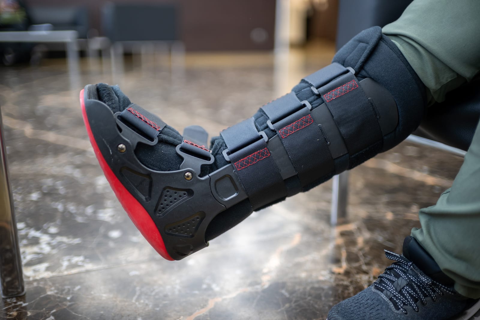
Appropriate immobilisation is a cornerstone of early Grade II management, with structured lateral support more important than for a Grade I injury.
Compression bandaging is appropriate for the immediate post-injury period to help control swelling but is insufficient as a sole stabilisation strategy. A semi-rigid lace-up ankle brace is one of the preferred early management options for most Grade II presentations, providing lateral stabilisation while permitting sagittal-plane motion and facilitating normal gait. Another alternative is a below-knee walking boot for patients with significant pain and functional limitation, or where the clinical picture warrants more complete off-loading of the lateral complex.
The duration of bracing or boot and the timing of progression from a walking boot to a functional brace depend on individual injury characteristics and recovery trajectory. Having the bracing type and duration directed by an orthopaedic specialist, rather than self-selected from a pharmacy shelf, gives the injury the best chance of healing without compromise. AVA Orthopaedics online consultations include bracing or boot recommendations tailored to the individual patient's injury grade, activity level and functional goals.
Physiotherapy and Rehabilitation: Structured Recovery With Clear Milestones
Physiotherapy-guided rehabilitation is the most important determinant of long-term outcome following a Grade II ankle ligament tear. A structured programme with clear progression criteria reduces the risk of re-injury and chronic instability.
Phase 1: Acute Management and Swelling Control, Days 1 to 7
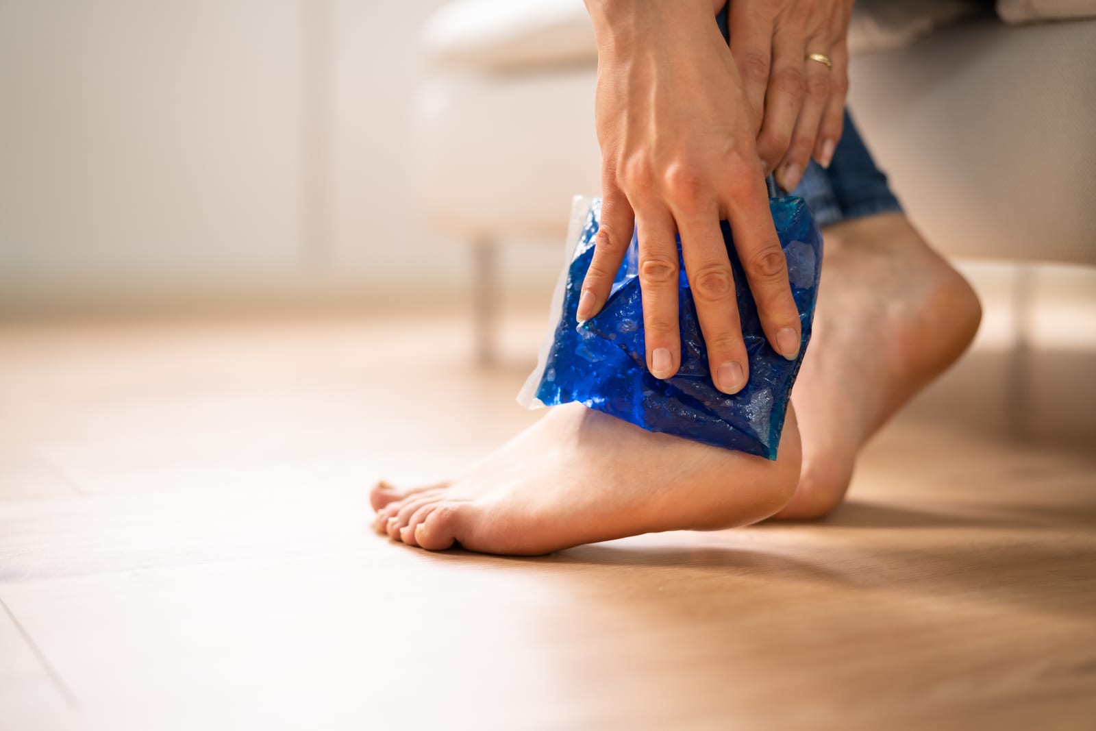
The PRICE protocol forms the basis of early management: protection, relative rest, ice, compression and elevation. HARM factors - heat, alcohol, running and massage - should be avoided in the first 72 hours. Gentle range-of-motion exercises within a pain-free range, including ankle alphabet movements and towel stretches, are introduced early. Weight-bearing progresses from partial to full within the brace as swelling and pain allow. Appropriate analgesia supports comfort throughout this phase.
Phase 2: Restore Range of Motion and Initiate Strengthening, Weeks 1 to 3
Progressive restoration of ankle dorsiflexion and plantarflexion range of motion is central to this phase. Peroneal strengthening begins with resisted eversion exercises using resistance bands and progresses to calf raises. Gastrocnemius and soleus stretching addresses any dorsiflexion restriction. Proprioceptive retraining begins on stable surfaces, starting with single-leg standing with eyes open and progressing to eyes closed.
Phase 3: Functional Strengthening and Proprioceptive Retraining, Weeks 3 to 6
Progressive single-leg strengthening exercises, step-ups, single-leg calf raises, and mini-squats build the dynamic stabiliser strength required for a return to sport. Proprioceptive training progresses to unstable surfaces, including balance board and wobble cushion work. Gait retraining normalises walking and jogging mechanics. Straight-line jogging is introduced when full pain-free weight-bearing and adequate range of motion have been achieved.
Phase 4: Sport-Specific Rehabilitation and Return-to-Sport Milestones, Weeks 6 to 10
Cutting, pivoting and reactive agility drills are introduced progressively alongside sport-specific movement patterns. Return to full competition requires meeting objective criteria: full, pain-free range of motion symmetrical with the uninjured side; peroneal strength symmetry of at least 90 per cent; single-leg hop test symmetry; and successful completion of sport-specific agility tasks without pain or apprehension. Prophylactic bracing for the first competitive season following return to sport may be recommended.
High Ankle Sprain Treatment: Recognising When the Injury Is Higher Than It Appears
The syndesmotic ligament complex, which binds the distal tibiofibular joint, is a distinct structure from the lateral ankle ligaments. A high ankle sprain occurs when external rotation of the foot on a fixed tibia stresses this complex, and it is particularly relevant in contact sports. In the context of a Grade II lateral ligament injury, a concurrent syndesmotic injury is an important possibility. The two can coexist, and a missed syndesmotic injury dramatically prolongs recovery.
Distinguishing features of a high ankle sprain include pain proximal to the lateral malleolus, a positive squeeze test, pain with external rotation stress and prolonged difficulty pushing off through the forefoot. High ankle sprain treatment principles differ significantly from those for lateral ligament management: longer protected weight-bearing, strict activity restriction, and, in cases of complete syndesmotic disruption with diastasis, surgical stabilisation is required.
Accessing Specialist Orthopaedic Care From Anywhere in Australia

Many patients with a Grade II ankle ligament tear are seen in an emergency department or by a GP and discharged without a clear specialist management plan. That gap in the pathway is where recovery is often compromised.
AVA Orthopaedics provides specialist online orthopaedic consultations via secure video, delivering the same standard of clinical assessment and management planning as an in-person appointment. For patients in regional, rural and remote areas of Australia where local orthopaedic services may be limited, or patients with busy lives juggling work and their own kids' activities, this means that specialist input can be obtained without delay.
An AVA Orthopaedics online consultation for a Grade II ankle injury includes a structured clinical history and imaging review, specific bracing or moonboot and load management recommendations, a physiotherapy referral and follow-up review to monitor progress and adjust the management approach as required.
Book an Online Orthopaedic Assessment for Specialist Advice, Treatment Direction and Clarity
A Grade II ankle ligament tear creates genuine clinical uncertainty: has a fracture been excluded, is the current brace appropriate, and what does the return-to-sport pathway actually look like? A consultation with an orthopaedic specialist at AVA Orthopaedics answers those questions clearly and precisely.
Before your appointment, having any existing imaging reports and images available, along with your GP or emergency department discharge summary if applicable, a clear account of how the injury occurred and your current symptoms and functional limitations, ensures the consultation is as productive as possible. Appointments are available via secure video from anywhere in Australia, often on the same day or the next.